Brief Description
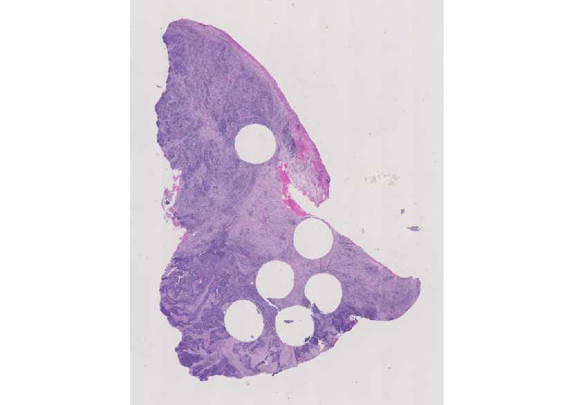
During the scan phase, WSI systems usually skip blank areas where tissue is absent in order to reduce scan time and file size. The purpose of the whole slide tissue coverage
836 test is to demonstrate that all of the tissue specimen on the glass slide is included in the digital image file. Tuis page is dedicated to methods for measuring WSI tissue coverage.
Status
Under Development: Recruiting authors and reviewers
Ready for Science-Sharing Seminar: See page with session proposal
Ready for Drafting for White Paper: See page with authors and outline
Published
Authors
Chronicle authors and contributors and their contributions/role (author, editor, date). Please include links to NCIPhub profile pages to facilitate messages to page contributors.
Overview
Keep this section about one page.
Why Important
“What you would tell your Mom.
Related System Components
System-level is an appropriate response here.
Materials and Methods
Keep this high level. Show pictures and figures. For details, add new wiki pages and link to new pages: wiki-internal or wiki-external.
How related to tissue and the pathologist
Does this impact the pathologist? How?
WSIWG Presentations and related Wiki pages
Document the discussions (when, where, what) and provide links to related slides.
- Oct. 12, 2015: Uwe Horchner, Joachim Schmid, and Anindya Sarkar, “Tissue Detection,” presentation given at the joint session of the WSI WG and DPA at Pathology Visions, Boston, MA. Link
- Oct. 12, 2015: Tyler Keay (Omnyx), “Whole Slide Tissue Detection,” presentation given at the joint session of the WSI WG and DPA at Pathology Visions, Boston, MA. Link
References
Format: authors (year), title, journal, volume, page, and then link to listing on journal website and the National Library of Medicine.